Pioneering experiment paves way to new imaging method for single molecules
An international research team including the QOQI team has succeeded for the first time in using X-rays for an imaging technique that exploits a special quantum property of light. As the researchers write in the journal Physical Review Letters, this technique could enable imaging of non-crystallized macromolecules.
The research team used the extremely short and very intense X-ray pulses of the European XFEL free-electron laser in Hamburg, Germany, to generate fluorescence photons that arrive at the detector almost simultaneously – in a window of less than a femtosecond (quadrillionth of a second). By calculating photon-photon correlations in the fluorescence of the irradiated copper atoms, it was possible to obtain an image of the light source.
At the atomic level, the structures of materials and macromolecules are usually determined by X-ray crystallography. While this technique relies on coherent X-ray diffraction, incoherent processes such as fluorescence emission can also occur and even dominate when X-rays are scattered, although they make no useful contribution to the diffraction measurement. Instead, they add a featureless fog or background to the measured data.
As early as the 1950s, however, two British astronomers demonstrated that it was possible to extract structural information from such light from self-luminous sources – in their case, light from stars. Robert Hanbury Brown and Richard Twiss‘ method, called intensity interferometry, opened a new door to understanding light and founded the field of quantum optics.
Researchers from QOQI, the Max Planck Institute for Structure and Dynamics of Matter and the German Electron Synchrotron (DESY) had recently proposed to apply intensity interferometry to atomic-resolution imaging using X-ray fluorescence [see A. Classen et al., PRL 119, 053401 (2017)]. The challenge in extending this idea to X-rays is that the photon coherence time, which determines the available time interval for photon-photon correlations, is extremely short. It is determined by the excited atom’s radiation decay time, which is about 0.6 femtoseconds for copper atoms.
Now, the group, together with researchers from Uppsala University and the European XFEL, has overcome this challenge: It used XFEL pulses with a duration of femtoseconds to trigger X-ray fluorescence photons within the coherence time. The team created a source of two fluorescent spots in a copper foil and measured the fluorescence on a one-million-pixel detector eight meters away.
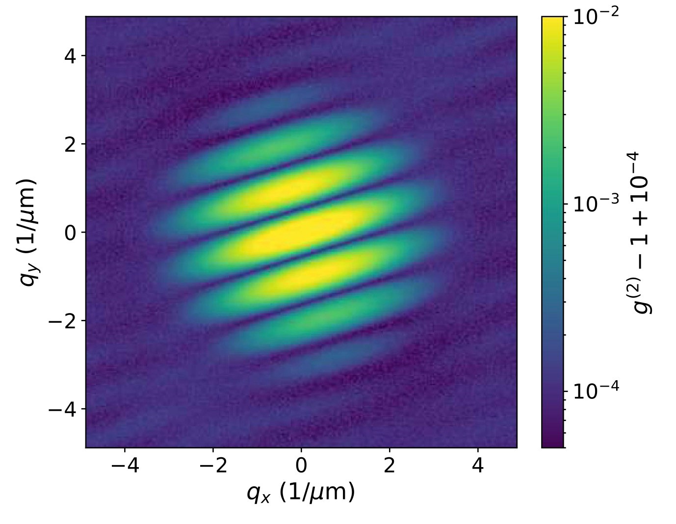
For each illumination pulse, only about 5,000 photons were detected by the detector, and the cumulative total over 58 million pulses yielded only an inconspicuous uniform distribution. However, if the scientists summed the photon-photon correlations over all detector images instead, a fringe pattern emerged. This fringe pattern was then analyzed like a coherent wavefield to reconstruct an image of the fluorescence source consisting of two well-separated spots of illumination.
The researchers now hope to combine the novel method with conventional X-ray diffraction to image individual molecules. In doing so, the element-specific fluorescent light can reveal substructures specific to certain atoms and even certain chemical states. This could help to decipher the functioning of important enzymes involved in photosynthesis, for example.
Publication: Imaging via Correlation of X-Ray Fluorescence Photons, Fabian Trost et al., Phys. Rev. Lett. 130, 173201 (2023)
Physics Viewpoint: Bringing Interferometric Imaging into the X-Ray Regime